Cross Section Of A Compact Bone
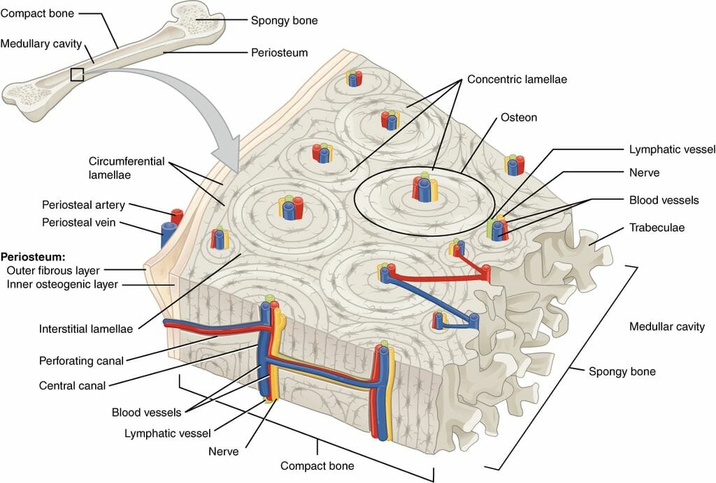
Cross Section Of A Compact Bone. The cross section of this circular cylinder is a circle. Compact bone is the outer layer and the spongy bone forms the inner layer. Skull bone is a flat bone. ( ) each osteon has a central haversian canal , running parallel to long axis of bone. In long bones, as you move from the outer cortical compact bone to the inner medullary cavity, the bone transitions to spongy bone. Two types of bone tissues in cross section of a long bone : Within each lamella, collagen is mixed with inorganic minerals like magnesium, calcium and phosphorus and layered around a haversian canal. The spongy and compact bone tissue in the cross section of a skull bone. The bottom sections of the spine are important when it comes to. Such roundish unit is called osteon.
Also called cortical bone, the compact variety usually features a haversian system, or cylindrical unit within the structure. Such roundish unit is called osteon. Two types of bone tissues in cross section of a long bone : As the names suggest compact bone looks compact and the spongy bone looks like sponges.
By printing out this quiz and taking it with pen and paper creates for a good variation to only playing it online.
Long, short, sesamoid (like the knee cap) , irregular, and flat. We can see there are two layers of compact bone here. This slide contains a section of dried compact bone. Vector illustration scheme of bone cross section. The spongy and compact bone tissue in the cross section of a skull bone. Compact bone is the heaviest, hardest type of bone. It consists of two layers; Also called cortical bone, the compact variety usually features a haversian system, or cylindrical unit within the structure. To the left is muscle tissue, and to the right Such roundish unit is called osteon. Cross section of spongy bone :
The compact bone is made up of osteon. Also called cortical bone, the compact variety usually features a haversian system, or cylindrical unit within the structure. Sketch and label of a cross section of a long bone / compact bone, makes up the dense material in a long section of a bone.
This is known as the periosteum.
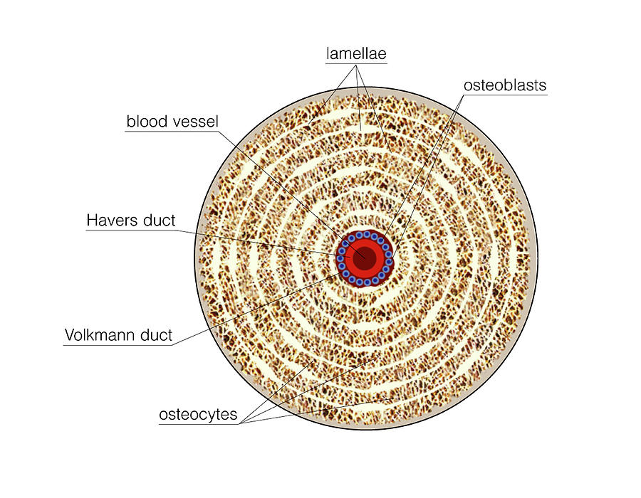
Long, short, sesamoid (like the knee cap) , irregular, and flat. The large dark spots are passages for blood vessels and nerves. Table 1 portion of bone width of the whole cross section width of the marrow section width of the compact bone section % of bone cross section that is compact bone chicken leg 1.0 cm 0.8 cm 0.2 cm 20% beef leg 4.5 cm 2.7 cm 1.8 cm 40% 3. Find the perfect bone cross section stock photos and editorial news pictures from getty images. Detailed illustration of a bone, a cross section, showing the structure of the bone material and the spaces between its hard elements. They conduct blood vessels, lymphatics, and nerves throughout the bone. Vector illustration scheme of bone cross section. Labeling portions of a long bone. A cross section of a compact bone shows concentric circles called lamellae. Within each lamella, collagen is mixed with inorganic minerals like magnesium, calcium and phosphorus and layered around a haversian canal. Compact bone is the heaviest, hardest type of bone. The compact bone is made up of osteon. The cross section of this circular cylinder is a circle. Compact bone cross section courtesy:
Cancellous bones, compact bone, cortical bone, diaphyses, haversian canal, lamella, marrow cavity, osseous tissue, osteons. The cross section of a rectangular pyramid is a rectangle. Skull bone is a flat bone. We can see there are two layers of compact bone here.

The large dark spots are passages for blood vessels and nerves.
A cross section of a human long bone. Some, mostly older, compact bone is remodelled to form these haversian systems (or osteons).the osteocytes sit in their lacunae in concentric rings around a central haversian canal (which runs longitudinally).the osteocytes are arranged in concentric rings of bone matrix called lamellae (little plates), and their processes run in interconnecting canaliculi. The compact bone is made up of osteon. In each osteon, the lamellae are arranged around a central haversian canal that houses nerves and blood. To the left is muscle tissue, and to the right Cross section of a femur bone showing the anatomical structure including cancellous bone and marrow. Bone markings the surface features of bones vary considerably, depending on the function and location in the body. Cancellous bones, compact bone, cortical bone, diaphyses, haversian canal, lamella, marrow cavity, osseous tissue, osteons. Preparation of bone cross sections with implants from d12oja0ew7x0i8.cloudfront.net browse 53 bone marrow cross section stock photos and images available, or search for bone cross section or bone cells to find more great stock photos and pictures. By printing out this quiz and taking it with pen and paper creates for a good variation to only playing it online.
By printing out this quiz and taking it with pen and paper creates for a good variation to only playing it online cross section of a bone. We can see there are two layers of compact bone here.

Posting Komentar untuk "Cross Section Of A Compact Bone"